The 49th Annual Meeting of Society for the Study of Reproduction (SSR) took place on 16–20 July 2016 in San Diego, USA.
This scientific meeting covers research in reproductive biology and fertility and was attended by over 900 delegates.
The highlights of the meeting included: New methods for diagnosis and management of endometriosis; miRNAs and endometriosis: potential biomarkers and targets; Life in HD
New approaches for diagnosis and management of endometriosis
During the conference novel and emerging technologies were showcased, which could have important implications in endometriosis. Research led by Dr Andrea Braundmeier-Fleming from Southern Illinois University presented a novel approach to endometriosis diagnosis by investigating the urogenital micro-biome.

Diverse E.coli by Mattosaurus (Own work) [Public domain], via Wikimedia Commons
The human body is colonised by complex communities of microbes in different micro-environments that vary across health and disease throughout the lifespan. The micro-biome is the collective genome of the microorganisms that colonise a given environmental niche.
The group hypothesised that if changes in inflammation in the female reproductive tract could alter the micro-biome then assessing the micro-biome in patients with and without endometriosis could provide a new diagnostic tool. The group recruited patients undergoing laparoscopic surgery for pelvic pain with suspected or previously diagnosed endometriosis and control patients undergoing surgery for benign uterine or ovarian conditions. They assessed vaginal swabs collected pre-surgically and during follow up from surgery and assessed microbial communities using16S ribosomal RNA (rRNA) sequencing, a common sequencing method used to identify and compare populations of different bacteria present within a given sample. Their preliminary results suggest that the urogenital micro-biome from patients with endometriosis is unique when compared to control patients concluding:
The data from this study indicates a specific urogenital micro-biome signature associated with endometriosis which could be utilised as a non-invasive diagnostic tool for endometriosis
Another approach highlighted was the use of transcriptomics to understand the impact of disease on fertility. Professor Carlos Simon from the University of Valencia has led the development of the endometrial receptivity array an approach that has examined the transcriptional profile of endometrial tissue samples from women to assay endometrial receptivity or ‘readiness’ for pregnancy. This gene expression data was used to develop a predictive model to determine the window of implantation in women [1,2].
The recent developments presented at the SSR meeting highlight the improved, version 2.0 model. This new model has been retrained based on a larger patient cohort consisting of endometrial samples from 600 patients including those that were receptive and went on to become pregnant. The expansion of this dataset and improvement of the predictive model may allow the researchers to identify and stratify responses of patients and allow different states of health and disease to be taken into account when assessing fertility. They explained how the technology could be used in patients to identify incidence of non-receptive endometrium, which may be associated with a disease-specific transcriptional signature. In the future this could allow for more targeted management of fertility assessment in endometriosis patients and may aid the diagnosis of endometriosis in patients presenting with sub/infertility.
Micro RNAs: potential biomarkers and targets
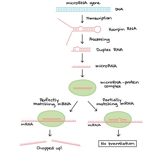
MicroRNAs (miRNA or miRs) can control cell function through post-transcriptional regulation of gene expression. MiRs are reported to be dysregulated in endometriosis tissue which may contribute to the pathophysiology of the disease.

Diagram of where miRNAs come from and how they regulate targets. Image from https://www.khanacademy.org/science/biology/gene-regulation/gene-regulation-in-eukaryotes/a/regulation-after-transcription. Image modified from “miRNA biogenesis,” by Narayanese, CC BY-SA 3.0. The modified image is licensed under a CC BY-SA 3.0 license.
In the focus sessions on ‘Normal Uterine Development, Physiology, and Dysfunction’ a number of researchers presented work related to miRNAs and endometriosis. Dr Niraj Joshi from Michigan State University examined the role of miR-451 in contributing to cell proliferation in endometriosis through regulation of its putative target YWHAZ (14.3.3zeta – promotes cell proliferation and inhibits apoptosis). Presenting work from a baboon model of endometriosis as well as studies using immortalised human endometriotic epithelial cells (12Z cells), Dr Joshi demonstrated that decreased expression of miR-451 in endometriosis alters the expression of cell cycle genes which can lead to the promotion of cell proliferation. They reasoned that this aberrant proliferation may contribute to the persistence of endometriotic lesions and the progression of the disease. Notably, when the group over-expressed miR-451 in vitro this inhibited G1/S transition in cells. Thus changes in miR-451 may control proliferation in endometriosis suggesting:
a potential role for miR-451 as a novel [approach] for targeted therapy for endometriosis
Presenting evidence from a mouse model Dr Warren Nothnick from the University of Kansas demonstrated that the miR-451 target macrophage migration inhibitory factor (MIF) can reduce endometriosis lesion size. Dr Nothnick also suggested that miR-451 has the potential to be used as a therapeutic target in endometriosis as it targets proteins that play a role in cell proliferation, invasion and inflammation, key processes in endometriosis. Furthermore Dr Nothnick highlighted that expression of miR-451 is detectable in plasma and therefore could have potential utility as a biomarker for endometriosis.
Life in HD

Eric Betzig, PhD Janelia Group Leader Howard Hughes Medical Institute
Optical microscopy has been critical to our understanding of cellular physiology. Nobel laureate Eric Betzig (HHMI, Janelia research campus) presented the highlight of the conference in his plenary lecture “Imaging Life at High Spatiotemporal Resolution”. This state-of-the-Art symposium lecture explained the breakthroughs in microscopy techniques that have driven forward research in recent years. However Dr Betzig began by outlining the limitations of microscopy methods to answer the questions of biology claiming
We have never seen cells in their natural state
Betzig’s research has sought to address this very problem, striving to develop an ‘optical microscope that could look at living cells with the resolution of an electron microscope.’ The discovery of photo-activatable GFP allowed Betzig to develop photo-activated localisation microscopy, or PALM as it is more commonly known, building the first PALM microscope in the living room of collaborator Harald Hess. PALM utilised a method of controlling excitation of fluorescent proteins with pulses of light. PALM microscopy was named Nature Methods technique of the year 2008 and Betzig later shared the Nobel Prize in Chemistry in 2014 for his role in the development of super-resolved fluorescence microscopy. This breakthrough, harnessed a new way of imaging by activating individual fluorophores over different discrete points in space and time to create an image with incredible resolution (nanometer scale). ‘This makes it possible to track processes occurring inside living cells’. Betzig went on to describe the latest advances in microscopy and imaging that go beyond his initial work but warned that the latest techniques are pushing the boundaries of what we can process
Lattice sheet microscopy is the future if we can find a way to handle the data
PALM and similar technologies are now becoming commonplace in research environments and these new approaches are now being used to better understand cell biology and provide new opportunities for insights into disease physiology.
Summary
The SSR meeting detailed the latest approaches and techniques that will help to inform current and future research into endometriosis. The fundamental research presented emphasised the possibilities of new methods for non-invasive diagnosis and therapeutic targeting of endometriosis. These advances highlight the benefit of collaborative interactions between scientists and clinicians to ensure the best chances of translating cutting-edge research into clinical practice.
References
- Díaz-Gimeno P, Ruiz-Alonso M, Blesa D, Bosch N, Martínez-Conejero J, Alamá P, et al. The accuracy and reproducibility of the endometrial receptivity array is superior to histology as a diagnostic method for endometrial receptivity. Fertility and Sterility 2013;99(2):508-517.
- Díaz-Gimeno P, Horcajadas J, Martínez-Conejero J, Esteban F, Alamá P, Pellicer A, et al. A genomic diagnostic tool for human endometrial receptivity based on the transcriptomic signature. Fertility and Sterility 2011;95(1):50-60.e15.
About the author

Douglas Gibson
University of Edinburgh
Douglas Gibson is a postdoctoral research fellow in the MRC Centre for Inflammation Research at the University of Edinburgh, United Kingdom. His research is focused on determining the impact of local steroid signalling in the regulation of endometrial function and in the pathogenesis of endometriosis. His research has shed new light on the role of tissue metabolism of steroids in regulation of endometrial tissues and offer insights into regulation of stromal differentiation (decidualisation), immune cell function, and vascular remodelling.

