
The first David Healy Award for best Scientific Abstract was presented to Mohamed Ibrahim at the closing ceremony of the 12th World Congress on Endometriosis for his work on pale cells diapedesis in the endometrial-myometrial junctional zone, providing new insights on the pathogenesis of adenomyosis.
Dr Ibrahim and his colleagues from the Charite University of Medicine in Germany conducted an ultrastructural study of the junctional zone (JZ) in adenomyosis (AM) to elucidate the enigma of the pathogenesis of AM and to search for microtrauma evidence in the JZ.
Methods
Twenty-four uteri from premenopausal women underwent laparoscopically-assisted vaginal hysterectomy were included and assigned to AM group (12 uteri) and non-AM group (n=12). Biopsies were obtained from the JZ of the anterior, posterior and fundus of the uterus at the midline, being the most prevalent sites of AM occurrence.
The JZ biopsies at the fundo-cornual raphe were studied using the transmission electron microscopy (TEM) and stained with van Gieson stain for collagen fibers. In addition, immunohistochemistry staining for E-cadherin, Transforming Growth Factor Beta Receptors (TGFβR) 1, 2 and 3 were performed.
Results
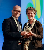
WES President, Linda Giudice, presents Mohamed Ibrahim with his award at the WCE2014 Closing Ceremony
TEM could show the so-named Pale cells, found eccentrically in the basal endometrial glands of AM. In different stages (partial/total detachment-thinning out-early diapedesis-late diapedesis), we could document different localisation of the Pale cells inside and outside the basal glands. They lacked desmosomal connection to the neighbouring epithelial cells. Ultramicrorupture of the basal lamina of these glands opposing these cells and abnormal nuclear infolding of the glandular epithelial cells were evident. The inner myometrial muscle fibers lost their parallelism, mimicking the outer myometrium in AM. No significant difference in E-cadherin, TGFβR1 or 2 expressions between both groups could be detected. Only TGFβR3 was significantly higher expressed in AM than non-AM.
Conclusion
Pale cells are speculated to be endometrial epithelial stem cells which can actively migrate into the myometrium – eased by lack of desmosomes – where they transform into AM, in a process might be mediated by TGFβR3. Ultramicrorupture at the JZ in AM is evident.
Shortlist for the Award
In addition to Dr Ibrahim, the following were short-listed for the David Healy Award 2014:
- Hans Albertsen | Salt Lake City (USA)
for his presentation on “GENOMIC REARRANGEMENTS (COPY NUMBER VARIANTS) MAY PLAY A ROLE IN THE PATHOGENESIS OF ENDOMETRIOSIS” - Erin Greaves | Edinburgh (UK)
for her presentation on “ESTROGEN RECEPTOR SUBTYPES MODULATE HOW NERVE FIBRES INFLUENCE BLOOD VESSELS AND MACROPHAGES IN PERITONEAL ENDOMETRIOSIS” - Louis Marcellin | Paris (France)
for his presentation on “EPIGENETIC AND GENOMIC ANALYSES OF HUMAN FETAL MEMBRANES FROM PREGNANT ENDOMETRIOSIS-AFFECTED WOMEN” - Vicky Young | Edinburgh (UK)
for her presentation on “METABOLIC REPROGRAMMING OF THE PERITONEAL MESOTHELIUM BY TGF-Β MAY PROMOTE ENDOMETRIOSIS LESION DEVELOPMENT”